Hi All
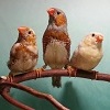
The strangest thing happened this morning, I was busy feeding my Canaries and they were all around me, some even landing and sitting on my arm when a Hen just fell over stone dead. I could not believe it, she was 100% one minute and dead the next. I picked her up and she was warm but as I said, dead. Do you think it was a heart attack?, I am stunned.
Steve
Heart Attack?
-
steve

- Flirty Bird

- Posts: 215
- Joined: Tue Nov 18, 2014 1:10 pm
- Location: South Africa
-
lovezebs

- Mod Extraordinaire

- Posts: 18214
- Joined: Sun Dec 15, 2013 11:51 am
- Location: Calgary Alberta Canada
Re: Heart Attack?
steve
I am so sorry to hear about your Canary girl.
Apparantly birds can have strokes, I am told, so I would imagine that a heart attack is possible as well. Perhaps she had some sort of a coronary abnormality, and with the excitement of being fed, maybe something just gave out.
Such a shame, the poor little thing probably didn't even know what hit her. At least it was quick and I am sure painless .
If you had an avian vet available, maybe he could perform an autopsy and be able to give you the reason of death.
Once again, I am sorry about your little bird.
~Elana~
~Elana~
Linnies~ Canaries ~ Zebras ~ Societies ~ Gouldians ~ Orange Cheeks ~ Shaft Tails ~ Strawberries ~ Red Cheek Cordon Bleu ~ Goldbreasts ~ Red Brows ~ Owls ~ Budgies ~ Diamond Firetails ~ Javas ~ Forbes Parrot Finches ~
Linnies~ Canaries ~ Zebras ~ Societies ~ Gouldians ~ Orange Cheeks ~ Shaft Tails ~ Strawberries ~ Red Cheek Cordon Bleu ~ Goldbreasts ~ Red Brows ~ Owls ~ Budgies ~ Diamond Firetails ~ Javas ~ Forbes Parrot Finches ~
-
steve
- Flirty Bird
- Posts: 215
- Joined: Tue Nov 18, 2014 1:10 pm
- Location: South Africa
Re: Heart Attack?
lovezebs
I went to the Vet Hospital but there was no Avian Vet there. I think this time of year is not ideal for services with everybody going on leave etc.
Steve
I went to the Vet Hospital but there was no Avian Vet there. I think this time of year is not ideal for services with everybody going on leave etc.
Steve
-
Jen

- Weaning

- Posts: 1452
- Joined: Thu Dec 05, 2013 9:02 pm
- Location: Beaumont, TX
Re: Heart Attack?
I'm so sorry about your hen. I also lost a hen suddenly this week. She was not as lucky as yours and took a long time to die. It is heart breaking to lose something we love. So sorry for your loss.
Jenny
Gouldians, Red Cheek Cordon Blue family, Gold Breasted Waxbills, Fire Finches, Owl finches, Yellow Face & Red Face Star Finches, Lavender Finches, Society Finches, Canary,Rosey Bourke, Scarlet Chested Grasskeets, Cockatiels, too many Guineas, Izzy my 16 year old cute doggie dog, two spoiled kitties!
-
cindy
- Bird Brain

- Posts: 18754
- Joined: Wed Jul 22, 2009 8:33 pm
- Location: west central Florida
Re: Heart Attack?
There can be many reasons for a sudden death, without a necropsy done it is uncertain what caused your bird's passing. So sorry for your loss.
Zebra, Gouldians, Java, CBM Shaft tail & Grasskeets
~ My Facebook groups ~
*Finchaholics ~ finches, hookbills, softbills & canaries are welcome here!
discussions regarding species, housing, breeding, preventatives, treatments
*Birdaholics ~ Avian Classified Ads Only
-
Sally
- Mod Extraordinaire
- Posts: 17929
- Joined: Thu Mar 29, 2007 11:55 pm
- Location: DFW, Texas
Re: Heart Attack?
So sorry for your loss. We often have no idea what went wrong.
-
fredbernie
- Hatchling

- Posts: 34
- Joined: Thu Dec 19, 2013 12:42 pm
Re: Heart Attack?
Birds of the Passerine, esp canaries, may become easy victims of stroke or heart failure. They can be knocked out even without any sign of fear or over-anxiety to trigger a stroke or heart attack. A friend of mine has a female who died suddenly from a stroke while sitting on eggs. Another one lost a male while it was standing and singing strongly on perch, literally singing then dropped dead. Normally, they are inherited diseases, so try to avoid breeding these birds and their descendants.
You may wish to have a look at the rear end of your bird, pay attention to the oil gland. If that is swollen and appears to have white substance inside, then your bird has suffered a stroke. If it is not then it seems to be a heart failure.
You may wish to have a look at the rear end of your bird, pay attention to the oil gland. If that is swollen and appears to have white substance inside, then your bird has suffered a stroke. If it is not then it seems to be a heart failure.
-
MiaCarter
- Molting

- Posts: 3528
- Joined: Wed Apr 30, 2014 1:36 pm
- Location: SW Florida
Re: Heart Attack?
fredbernie -- You wrote:
You may wish to have a look at the rear end of your bird, pay attention to the oil gland. If that is swollen and appears to have white substance inside, then your bird has suffered a stroke. If it is not then it seems to be a heart failure.
Are you referring to the preen gland?
I've heard something about this in passing. Do you know how is this an indicator of a stroke? How the stroke results in a change in the gland?
steve -- I'm so sorry you lost your girl.
At least it was quick and she didn't suffer. That's something, I guess.
They can absolutely suffer from strokes and heart attacks, just like humans, so very possible, as the others said.
If you would like, you could have her autopsied (it's actually called a necropsy in animals.) You would need to freeze her immediately, though, to halt decomposition. This is a service that's available at most veterinary clinics. It doesn't always result in an answer, but it may be worth a try if you bred this hen and need the insight.
Humum to....
13 Zebra Finches....and 2 squeeps!
3 Society Finches
6 Gouldians
1 Weaver
1 Pintail Whydah
2 Cockatiels
2 Parakeets
....along with 1 MinPin, 1 Pug, 1 JRT, 1 Yorkie, 2 Chihuahuas and 15 cats.


www.PetFinchFacts.com
13 Zebra Finches....and 2 squeeps!
3 Society Finches
6 Gouldians
1 Weaver
1 Pintail Whydah
2 Cockatiels
2 Parakeets
....along with 1 MinPin, 1 Pug, 1 JRT, 1 Yorkie, 2 Chihuahuas and 15 cats.
www.PetFinchFacts.com
-
cindy
- Bird Brain
- Posts: 18754
- Joined: Wed Jul 22, 2009 8:33 pm
- Location: west central Florida
Re: Heart Attack?
generally if there is an issue with a preen gland it is associated with a lack of vitamin A
http://www.exoticpetvet.net/avian/uropygial.html
"Diseases of the Uropygial Gland
Summary: The uropygial gland, when present, is a bilobed holocrinc gland with secretions that perform several functions in birds, including waterproofing (although not essential for it), manufacturing vitamin D precursors, keeping the skin, feathers, and bill supple, and performing antibacterial function. Many problems with this gland are secondary to hypovitaminosis A and and be corrected by moist hot packing, correcting the diet, and treating any secondary infections. Although impaction is often mentioned in the literature, this seems to not commonly occur. Diagnostics such as cytology, biopsy, aspiration, microbiology and histopathology may be used to procure a diagnosis. Surgery should only be attempted if medical management has been unrewarding, or if a tumor, abscess or rupture is present. Surgery requires excellent hemostasis and precise surgical technique.
Introduction
The uropygial gland, also called the preen gland or oil gland, is often overlooked during routine physical examination of birds. The avian practitioner should become familiar with the normal appearance of this gland (when present) to be able to identify abnormal glands. Most problems with the uropygial gland can be corrected by dietary and medical management, or rarely, surgery may be required. Not all species of birds have uropygial glands, and it is important to be knowledgeable about which species do, and which don't.
Anatomy and Physiology
The uropygial gland is a bilobed holocrinc gland. It is the principle cutaneous gland of birds. It is present in most species of bird, and it is relatively large in some aquatic species. It is absent in other species, including the ostrich, emu, cassowary, bustard, frogmouth, many pigeons, many woodpeckers and certain species of psittacines.1 Among the psittacine species that do not possess a uropygial gland are the hyacinth macaw, Anodorhynchus hyacinthinus , the Lear's macaw, Anodorhynchus leari, and the Spix's macaw, Cyanopsitta spixii. All of the parrots in the genus Amazona also do not posses a uropygial gland. The gland is present in the other psittacine species. The gland is also present in canaries and most finches.
The uropygial gland, when present, lies on the mid-line dorsally on the trunk in the rump area above the levator muscles of the pygostyle. In domestic fowl, the gland is drained by a pair of ducts. Each duct drains one lobe and each duct opens into a single, narrow nipple-like papilla. Other species have up to eighteen orifices.1 There are no feathers normally on the skin over the gland. There is, however, a tuft of down feathers at the tip of the papilla in most species, and this is called the uropygial wick. In many species of bird, the tail usually flexes laterally each time the bird reaches around to contact the gland and the wick.2
The gland secretion is complex and consists of a combination of extruded cells, ester waxes, fatty acids, fat and sudanophilic secretory granules.1,2 The secretion is spread over the feathers during the act of preening. Waterproofing is considered to be one function of the secretion (although it is not necessary for it), and another function is the suppression of the growth of organisms on the skin. The secretion helps keep the feathers, beak, and scales supple. The secretion is odorous in the female and nestling hoopoe, and in the musk duck and petrels.1
The secretion from the uropygial gland also contains vitamin D precursors that are also spread over the feathers by preening. With exposure to the ultraviolet portion of sunlight, the secretions are converted to an active form, vitamin D3, which is then ingested with subsequent preening.3
New research shows that the bird eye sees light in the ultraviolet range, and the secretion from the uropygial gland may also play a role in the identification of the sex of a bird, and may be involved with individual identification of birds, as well.4 In primates, the lens acts as a yellow filter which cuts off light of wavelengths below 400nm and therefore renders ultraviolet radiation invisible. However in diurnal birds, the cornea and lens are optically clear and appear to transmit wavelengths down to about 350nm, thus rendering near ultraviolet radiation visible.1 The lens only absorbs those ultraviolet wavelengths that are not physiologically destructive.1
This is a green-cheeked conure with a chronically impacted uropygial gland. The channels that allow the secretion to wet the wick feather are occluded. Hot packing the area with a warm, wet cloth and gentle massage will allow the secretion to begin flowing again. The secretion is of normal color, viscosity and consistency, and is visible through the skin and gland wall.
Examination
It is very important that the avian practitioner visually examine the uropygial gland in all birds where present. The gland has a size and shape variation from species to species. For example, the uropygial gland is raised and somewhat heart-shaped in the African grey parrot, Psittacus erithacus erithacus, and in comparison to that of other species, such as the eclectus parrot, Eclectus roratus, it may appear enlarged by comparison. However, the uropygial gland is just a larger gland in the grey, which is a normal species variation. This author has performed many second opinions concerning the uropygial gland of greys, which were thought to be enlarged, impacted, infected, or abnormal, but in actuality, the gland was normal for a grey. Another species with a prominent uropygial gland is the Moluccan cockatoo, Cacatua moluccensis.
Abnormalities of the Uropygial Gland
The most common abnormality of the uropygial gland occurs from vitamin A deficiency.5 This may cause glandular metaplasia and hyperkeratosis. Birds on poor diets are likely to be vitamin A deficient, and in addition to blunting of the choanal papillae that is commonly seen with hypovitaminosis A, enlargement of the uropygial gland is a likely sequela, as well. Although there are common references in the avian literature concerning impaction of the uropygial gland, in this author's practice, this is very rarely observed. The gland normally appears somewhat swollen, which may be mistaken for disease. With hypovitaminosis A, a hyperkeratotic plug may form in the gland, which may be dislodged by gently massaging or milking the gland after moist hot compresses have been applied. Correction of the diet and perhaps an injection of parenteral vitamin A, will usually rectify the problem.3
Neoplasia of the uropygial gland may occur. Adenomas, squamous cell carcinomas, papillomas, and adenocarcinomas have all been reported.3,5 Neoplasms may have variable appearances, may be unilateral or bilateral, and they may superficially ulcerate.5
Infection may also occur in the uropygial gland. This may be secondary to hypovitaminosis A, immunosuppressive disease, such as that which occurs with Psittacine Beak and Feather Disease (PBFD), or trauma. However, in this author's experience, infectious adenitis is a rare occurrence. Bacterial or fungal adenitis does not often occur, and when it does, it is usually in PBFD positive birds. Infected glands may abscess.
Another condition of the uropygial gland has been observed by this author, but has not been described in the avian literature. Two obese cockatiels presented with the primary complaint of staining and a greasy appearance over the area of the uropygial gland and retrices. Examination of the gland showed swelling and upon massage of the gland, an excessive amount of the oily gland secretion leaked out through the wick. Both of these cockatiels were hens, very obese, on poor all-seed diets, and color mutations. Both birds responded to a weight loss program, dietary changes, and increased exercise. Once the hens reduced their weights to the normal range, the uropygial glands ceased producing excessive secretions.
Rupture of the uropygial gland has been reported in gentoo penguins and in free-living seabirds in Europe.3
Chronic dermatitis of the skin over and surrounding the uropygial gland may occur and may respond to appropriate medical therapy based on the cause.5
Diagnostic Methods
Diagnosis of disease of the uropygial gland will require taking a thorough history of the bird and evaluation of the diet. Cytologic examination of the gland secretion may be diagnostic. This can be performed by gently massaging or milking the gland and touching a glass slide to the wick to collect material. The secretion may be cultured for both aerobic and anaerobic bacteria. The gland can be biopsied or an aspiration may be performed and submitted for histopathology.3
Treatment
Treatment of disease of the uropygial gland will depend on the diagnosis. Most abnormal glands will be the result of hypovitaminosis A, and may be secondarily infected. These cases will respond to parenteral supplementation of vitamin A, correcting the diet, and hot-packing the area with moist heat in cooperative patients. Based on culture and sensitivity results, systemic antibiotic or antifungal therapy may be beneficial.3,5
Suspected tumors may require surgical excision. Surgical excision should also be considered when conservative medical management has not been effective, if impaction recurs, there is chronic, non-responsive infection, or if the gland has ruptured. If possible, it is best to excise the gland before it ruptures, as the resulting inflammation, cellulitis, scar tissue, or septicemia may prove debilitating or life threatening.3 Surgery should be considered as a last resort for conditions that can be managed medically, and should not be attempted for hyperkeratotic glands. In most birds, other than ducks, surgical removal of the uropygial gland does not appear to clinically affect the bird, however, in ducks, the glands excision will result in the duck losing its ability to waterproof the feathers.3
Surgical Procedure
Surgical excision of the uropygial gland should be performed under general anesthesia and with radiosurgery to maintain hemostasis and minimize damage to the retrices. A fusiform incision is made over the gland extending beyond the papillae. The gland is bilobed, and each lobe receives its blood supply from three blood vessels. The gland may attach to the deep areolar fascia over the synsacrum and caudally to the insertion point of the retrices. Care must be taken to avoid the blood supply to the retrices. The gland is bluntly dissected, and the blood vessels are identified and coagulated. The gland is gently dissected until it can be removed. The fascia should be closed with monofilament absorbable suture in a pattern to reduce tension to the skin. The skin is closed with a simple interrupted pattern.6,7 Absolute hemostasis must be maintained during surgery, or seeping of blood may occur post-operatively, once the bird recovers from the anesthesia and the blood pressure returns to normal. This may result in large hematoma formation and may be involved in dehiscence.
If the gland has ruptured, or if it is severely chronically infected, extensive debridement may preclude total skin closure, and the wound may need to heal by second intention. Dehiscence and damage to the retrices may occur in these cases.6,7 A drain may be placed ventral to an infected, ruptured gland, and it may remain in place for three days. Some birds may require a collar to prevent post-surgical plucking, removal of sutures or drain.
Conclusions
The uropygial gland should be evaluated in every bird that has one. Observing the gland in many birds will give the practitioner a good idea of what they should normally look like, so that it will be easier to identify an abnormal one. Most problems with the uropygial gland are related to hypovitaminosis A and most respond well to medical therapy, including the application of moist heat, massage and appropriate therapeutics. Tumors, abscesses, and ulcers may require surgical intervention, and if at all possible, the gland should be surgically excised prior to its rupture. This gland produces secretions with several known functions, and as we learn more about birds, I suspect that we will find other functions, as well. More research needs to be performed concerning the anatomy and physiology of this gland so that we may better understand its structure and function.
By Dr Margaret Wissman D.V.M., D.A.B.V.P."
Zebra, Gouldians, Java, CBM Shaft tail & Grasskeets
~ My Facebook groups ~
*Finchaholics ~ finches, hookbills, softbills & canaries are welcome here!
discussions regarding species, housing, breeding, preventatives, treatments
*Birdaholics ~ Avian Classified Ads Only
-
cindy
- Bird Brain
- Posts: 18754
- Joined: Wed Jul 22, 2009 8:33 pm
- Location: west central Florida
Re: Heart Attack?
In order to have necropsy done do not freeze the body...freezing destroys cells...instead the body should be refrigerated in an air tight zip lock bag and brought to the vets asap
http://www.quakerville.com/qic/qnecrops.asp
"Body Preparation NEVER freeze a bird's body. This destroys tissue by crystallizing the fluids within. The sooner after death the body is sent to a lab, the greater the chances of discovering the exact cause of death. As soon as you discover a bird had died, place the body in an airtight plastic bag in a refrigerator. If possible, transport to the lab in an ice chest to retain the temperature. Bring along a thorough history of the bird's life, including age, length of time owned, size of cage, exercise routine, diet, contact with other birds, records of vet visits, and any unusual symptoms that may have been exhibited. Visual Inspection ... To help speed the results, the pathologist first studies the bird's case history for hidden clues. Items that may seem insignificant to you may prove quite useful in assisting the pathologist in determining the cause of death."
Zebra, Gouldians, Java, CBM Shaft tail & Grasskeets
~ My Facebook groups ~
*Finchaholics ~ finches, hookbills, softbills & canaries are welcome here!
discussions regarding species, housing, breeding, preventatives, treatments
*Birdaholics ~ Avian Classified Ads Only
-
MiaCarter
- Molting
- Posts: 3528
- Joined: Wed Apr 30, 2014 1:36 pm
- Location: SW Florida
Re: Heart Attack?
cindy - Thanks for that correction on refrigerating instead of freezing!
I was always told to freeze (but thinking back, in both cases where I was told to freeze, we were sending out-of-state. So perhaps that's the reason.)
But that totally makes sense. When water freezes into ice, it enlarges, thereby causing cell damage/rupture.
That was my understanding on the preen gland too.
I'm curious as to how a stroke could bring about a change in the gland.
Humum to....
13 Zebra Finches....and 2 squeeps!
3 Society Finches
6 Gouldians
1 Weaver
1 Pintail Whydah
2 Cockatiels
2 Parakeets
....along with 1 MinPin, 1 Pug, 1 JRT, 1 Yorkie, 2 Chihuahuas and 15 cats.
www.PetFinchFacts.com
13 Zebra Finches....and 2 squeeps!
3 Society Finches
6 Gouldians
1 Weaver
1 Pintail Whydah
2 Cockatiels
2 Parakeets
....along with 1 MinPin, 1 Pug, 1 JRT, 1 Yorkie, 2 Chihuahuas and 15 cats.
www.PetFinchFacts.com
-
cindy
- Bird Brain
- Posts: 18754
- Joined: Wed Jul 22, 2009 8:33 pm
- Location: west central Florida
Re: Heart Attack?
Stroke occurs within the brain,.... the preen gland is separate. The only true way to find out is necropsy.
The bird may have had something internal wrong that may have shortened her life, maybe something from births.
Steve, how old was she? A young bird is unlikely to have a stroke.
The bird may have had something internal wrong that may have shortened her life, maybe something from births.
Steve, how old was she? A young bird is unlikely to have a stroke.
Zebra, Gouldians, Java, CBM Shaft tail & Grasskeets
~ My Facebook groups ~
*Finchaholics ~ finches, hookbills, softbills & canaries are welcome here!
discussions regarding species, housing, breeding, preventatives, treatments
*Birdaholics ~ Avian Classified Ads Only
-
fredbernie
- Hatchling
- Posts: 34
- Joined: Thu Dec 19, 2013 12:42 pm
Re: Heart Attack?
Thanks cindy for the paper, I did read it before.
Abt the preen gland, I'm not sure how and why a stroke would have any effects on it. I also have doubt abt it at first, but I experienced it myself, and some breeders in my local club also told me abt it. I guess it is a very viable research subject for some avian physiologists. The common first-aid treatment for stroke that I've been using with certain success (11 out of 15 birds summing up from all local breeders recovered from stroke and some actually have success in breeding after that) is to gently squeeze the preen gland to extract all white substances as soon as a stroke is noticed. Then apply some eucalyptus oil on the feet and the preen gland, and keep the bird warm under the heat from an incandescent light bub. If the bird can recover, it would be after 48-72 hours. A proper caring program focusing on good diet and with plenty of room for exercise to regain control of the body would bring it to the full healthy condition after 8-12 weeks . Generally, the sooner the treatment takes place, the higher the chance for recovery. Some birds may die during the treatment cycle, and some may have stroke for a second time. So it is purely a common experience drawn from practice, not a scientifically proven method.
Abt the preen gland, I'm not sure how and why a stroke would have any effects on it. I also have doubt abt it at first, but I experienced it myself, and some breeders in my local club also told me abt it. I guess it is a very viable research subject for some avian physiologists. The common first-aid treatment for stroke that I've been using with certain success (11 out of 15 birds summing up from all local breeders recovered from stroke and some actually have success in breeding after that) is to gently squeeze the preen gland to extract all white substances as soon as a stroke is noticed. Then apply some eucalyptus oil on the feet and the preen gland, and keep the bird warm under the heat from an incandescent light bub. If the bird can recover, it would be after 48-72 hours. A proper caring program focusing on good diet and with plenty of room for exercise to regain control of the body would bring it to the full healthy condition after 8-12 weeks . Generally, the sooner the treatment takes place, the higher the chance for recovery. Some birds may die during the treatment cycle, and some may have stroke for a second time. So it is purely a common experience drawn from practice, not a scientifically proven method.
-
afinch
- 3 Eggs Laid

- Posts: 758
- Joined: Sat Apr 05, 2014 10:12 pm
Re: Heart Attack?
MiaCarter if you're sending "sample" for bacteriology/virology/metabolic testing, then freezing might be ok (or required). However, freezing will destroy tissue and lyse cells, making necropsy difficult and complicated. So it's a significant last resort trade off.
-
afinch
- 3 Eggs Laid
- Posts: 758
- Joined: Sat Apr 05, 2014 10:12 pm
Re: Heart Attack?
fredbernie sounds to me like it's the other way around, i.e sounds like a problem with the gland (infection?) may lead to other complications (stroke?) unless attended to, rather than the gland necessarily being an indicator of stroke. Especially if as you say draining the gland and treating the possible infection/inflammation decreases that risk of additional complications.